Written by Ella Roor
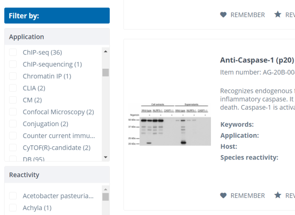
In our webshop you have the possibility to filter your product search by applications for which the respective products have been validated by the manufacturer. This allows you, for example, to quickly find an antibody you are looking for to use in immunohistochemistry or Western blotting. With our large product range, we also cover a wide range of applications, which is why we often use abbreviations for the respective application in the corresponding filter option for a better overview. However, these abbreviations are not always familiar. In this article we would like to give you an overview of some of our application abbreviations and explain a few selected methods in more detail.
| Application/Method | Abbreviation |
| Agglutination | AGG |
| Chromatin immunoprecipitation | ChIP |
| Dot Blot | DB |
| Ouchterlony double immunodiffusion | DD |
| Enzyme-linked Immunosorbent Assay | ELISA |
| Enzyme-linked ImmunoSpot Assay | ELISpot |
| Electrophoretic Mobility Shift Assay | EMSA |
| Functional Assay | FA |
| Flow Cytometry | FC |
| Fluorescence-linked Immunosorbent Assay | FLISA |
| Immunocytochemistry | ICC |
| Enzyme-linked Immunoelectro Diffusion Assay | ELIEDA |
| Electron microscopy | EM |
| Immunofluorescence | IF |
| Immunohistochemistry | IHC |
| Immunoprecipitation | IP |
| Lateral Flow Assay | LFA |
| Proximity Ligation Assay | PLA |
| Radioimmunoassay | RIA |
| Western Blot | WB |
Enzyme-linked Immunosorbent Assay (ELISA)
The ELISA is an immunological, antibody-based detection method for the examination of proteins (e.g. antibodies), viruses or small, biochemical molecules in different sample types (e.g. plasma, serum, urine). The method is characterised by high specificity and sensitivity, so that previously common techniques such as the radioimmunoassay (RIA) were gradually replaced by the ELISA [1, 2]. During the Corona pandemic, ELISAs had and have played an important role in the follow-up of SARS-CoV-2 virus infections, especially in asymptomatic infections. To test whether there was an infection with the corona virus, blood from the infected person was analysed by ELISA for antibodies specific to the SARS-CoV-2 virus [3]. A general distinction is made between four types of ELISA:
- Indirect ELISA
- Direct ELISA
- Sandwich-ELISA (direct/indirect)
- Competitive ELISA
The indirect ELISA uses an unlabelled primary antibody that specifically binds to the antigen in the sample. Subsequently, an enzyme-conjugated secondary antibody is used, which binds to the first antibody and enables the indirect detection of the antigen [4]. For the direct ELISA, the primary antibody is already conjugated with an enzyme, meaning that no secondary antibody is needed [4].
Compared to the first two variants, in the sandwich ELISA two antibodies bind to two different epitopes of the antigen. As a result, the antigen lies between the two antibodies, as in a sandwich. This type of ELISA is considered direct if the second antibody is conjugated with an enzyme. If this is not the case, a third antibody must be used, which is coupled to an enzyme and allows the detection of the antigen (indirect sandwich ELISA) [4].
The competitive ELISA is a special case: Here, a competing conjugated antigen is used instead of a second antibody. This antigen is structurally similar to the antigen from the sample and competes with it for the binding site of the detection antibody [5]. This results in an inversely proportional relationship between the concentration of the antigen to be detected and the signal strength (low signal strength = high antigen concentration in the sample) [5].
Regardless of the ELISA type, a colour substrate is added to the sample to detect the antigen. The enzymes conjugated with the antibodies convert the substrate, resulting in a faint yellow colour change. The intensity of the resulting colour change provides information about the concentration of the investigated antigen in the sample [1, 6].
Discover our selection of ELISA and Immunoassays
Flow Cytometry (FC)
Flow cytometry (FC) is an analytical method that enables the determination of the physical and chemical properties of cells. This technique is used, for example, in the detection of infections, in immunology or in cancer research. A flow cytometer that is used for this purpose, consists of three main systems: the fluidics, the optics and the electronics [7].
The principle behind FC is based on labelling cells with specific fluorescent dye-coupled antibodies or directly with cell- or organelle-specific dyes. The cells are then transported through a narrow channel where they are illuminated by a laser beam, so the electrons of the (fluorescent) dye are raised to an increased energy level. When the electrons fall back to their original energy level, they emit light, which is detected by the flow cytometer. The amount of emitted light is proportional to the amount of bound antibodies and thus to the cell number [7, 8]. The light scattering and diffraction that take place during this process allow the determination of the cell size and nature.
Some flow cytometers are also able to separate and fractionate cells. Once the cells have passed the laser beam, they are packed into small liquid droplets and have positive and negative charges. While moving through an electric field, the cells are directed into sterile collection vessels according to their charge. In this case, the method is called "fluorescence-activated cell sorting" (FACS). The collected data is then forwarded to a programme for evaluation and the results are displayed in diagrams (dot plots) [7-9].
Discover our selection of FC Antibodies
Immunohistochemistry (IHC)
Immunohistochemistry (IHC) is a method that is mainly used in oncological diagnostics. Tumours can be identified and classified by means of IHC, so that corresponding prognoses can be made and therapy approaches defined. Furthermore, IHC makes it possible to distinguish between healthy and diseased tissue. In addition to identifying tumours, IHC can also be used to detect infectious agents such as cytomegaloviruses, human papilloma viruses and herpes viruses [10,11].
The method uses antibodies which visualise the target proteins in the cell or tissue structures. The tissue must first be fixed for this, for which paraformaldehyde is generally used. For the subsequent detection of the target proteins, a distinction is made between two different methods: direct and indirect staining [12]. Direct staining uses primary antibodies coupled to a marker (fluorescent dye or enzyme). If the marker is a fluorescent dye (e.g. fluorescein, rhodamine or Texas Red), the protein under investigation is detected using a fluorescence microscope. In the case of enzymes, on the other hand, a chromogenic substrate is added to the sample, which is converted by the enzyme into a coloured reaction product, thus enabling antigen detection. Frequently used enzymes are in particular horseradish peroxidase (HRP) and alkaline phosphatase (AP) [12].
In contrast, indirect staining uses a non-conjugated primary antibody and a conjugated secondary antibody. The secondary antibody can in turn be conjugated with a dye or with an enzyme [11].
Discover our selection of IHC Antibodies
Lateral Flow Assay (LFA)
Lateral flow assays (LFA) are among the most commonly used biosensors in the field of analytical detection. They are characterized by being straightforward, inexpensive, and providing rapid results compared to other immunoassays (e.g., ELISA or Western blot). Moreover, no additional equipment or personnel are required to perform them [13]. Probably the best known LFA are pregnancy or covid-19 tests. But the assays are also used in other fields such as veterinary medicine, food safety or environmental sciences [14]. The LFA is used to detect analytes in liquids or complex mixtures (e.g., blood, urine) using a combination of thin-layer chromatography and immunoassay [14, 15].
Basically, all components required for an LFA are contained in the test cassette (Fig. 1). The test strip ("membrane") inside the test cassette consists of porous paper, which absorbs the sample and transports it within the paper with the aid of capillary forces [14].

Figure 1: Illustration of the basic structure of an LFA and the flow direction of the sample. An LFA generally consists of the following components: sample pad, conjugate release pad, a membrane with bound antibodies on test and control line and an adsorbent pad. The components are fixed on a backing card. After application of the sample, it moves from the sample to the adsorbent pad by capillary forces [14].
The sample is mixed with a solvent or running medium and applied to the sample pad, which filters the sample liquid so that transport is not impeded by solids within the liquid. The sample then moves through the "conjugate release pad", to which labeled primary antibodies have been immobilized. The sample fluid releases these antibodies, whereupon they bind to the antigen [14, 16]. Once the sample fluid has passed the conjugate release pad, it moves together with the conjugated antibody into the detection zone between the sample pad and the adsorbent pad. This zone is a porous membrane with immobilized test and control lines (Fig. 1). Antibodies directed against a different epitope of the analyte are immobilized on the test line. If the antigen is detectable in the sample, a distinct line appears, and the intensity of the staining is directly proportional to the antigen concentration [14, 16].
On the control line are antibodies immobilized, which specifically bind to excess, labeled primary antibodies and thus indicate the correct course of the test [2]. If only the test line is seen, this indicates a faulty test and the LFA should be repeated. To prevent reflux, there is also an "adsorbent pad" at the end of the test strip, which absorbs the excess amount of liquid [14, 16].
Discover our selection of LFA Antibodies
Western Blot (WB)
The Western Blot method celebrated its 40th birthday in 2019 [17]. Edwin Southern developed the first blot method, called Southern Blot, for the detection of DNA fragments in 1975. Based on this method, the Northern Blot (RNA) and Western Blot (proteins) techniques were introduced [18].
For a Western blot, the examined proteins must first be separated electrophoretically. For this purpose, various methods such as SDS-PAGE, native-PAGE, isoelectric focusing or 2D gel electrophoresis can be used. During a separation by SDS-PAGE, the folded proteins are denatured and linearised due to the usage of sodium dodecyl sulphate (SDS) and heating. In addition, the binding of the SDS to the proteins masks the protein intrinsic charge so that they have a permanent negative charge [19, 20]. During polyacrylamide gel electrophoresis (PAGE), proteins are separated according to their chain length, which is proportional to the molecular mass. Small molecules can pass through the gel matrix faster than large molecules [20].
This is followed by the actual "blotting". In this process, the separated proteins are transferred to a solid carrier membrane such as nitrocellulose, nylon or polyvinylidene difluoride (PVDF) by an electric field applied perpendicular to the gel [18]. The transferred proteins bind to the support material, which preserves the separation pattern and makes the proteins accessible for subsequent methods [19].
The most common subsequent method is the identification of the "blotted" proteins using specific antibodies (immunodetection). These can be directed either against the target protein itself or against a tag associated with the target protein (e.g. HA or GFP tag). To prevent or reduce non-specific binding of the antibodies, free binding sites on the membrane can be blocked, e.g. with the help of BSA. In addition, the carrier membranes should be washed with buffer solutions to remove antibodies that have not bound to the target protein. The primary antibody is detected with a secondary antibody directed against it, which is linked to a conjugate. This can be, for example, a dye or an enzyme [19].
The method has a high analytical sensitivity and specificity [21]. Compared to other analytical methods, the Western blot is characterised by the fact that a determination of the protein size, the protein content (when comparing different samples with each other) and the (semi-)quantification of the proteins are possible. Another advantage is the possibility to use different primary antibodies on the same carrier membrane, allowing the detection and comparison of different proteins from the same samples [19].
Discover our selection of WB Antibodies
Sources
[1] Rehm, H., Letzel, T. (2016). Antikörper und Aptamere. In: Der Experimentator: Proteinbiochemie/Proteomics. Experimentator. Springer Spektrum, Berlin, Heidelberg. https://doi.org/10.1007/978-3-662-48851-5_6
[2] https://flexikon.doccheck.com/de/ELISA 16.01.2023
[3] https://www.planet-wissen.de/natur/mikroorganismen/viren/elisa-test-112.html 16.01.2023
[4] https://elisa-kits.de/de/elisa_method 08.02.2023
[5] https://www.uniklinikum-saarland.de/de/einrichtungen/fachrichtungen/zellbiologie/seminar_zellbiologie_20192020/elisa/verschiedene_testformen/ kompetitiver_elisa 17.02.2023
[6] https://www.bionity.com/de/lexikon/Enzymelinked_Immunosorbent_Assay.html 16.01.2023
[7] https://at.vwr.com/cms/flow_cytometry 14.01.2023
[8] Oberle, V., Soßdorf, M., Lösche, W. (2010). Durchflusszytometrie. In: Pötzsch, B., Madlener, K. (eds) Hämostaseologie. Springer, Berlin, Heidelberg. https://doi.org/10.1007/978-3-642-01544-1_74
[9] https://www.laborjournal.de/rubric/methoden/methoden/m_v64.php?consent=1 18.01.2023
[10] https://ipa.med.ovgu.de/Krankenversorgung/Klinische+Pathologie/Immunhistologie.html#:~:text=Das%20wesentliche%20Anwendungsgebiet%20der%20Immunhistochemie,Ansprechen%20auf%20bestimmte%20Therapien%20genutzt.
[11] https://flexikon.doccheck.com/de/Immunhistochemie
[12] https://www.bionity.com/de/lexikon/Immunhistochemie.html 20.02.2023
[13] Jiang, X. & Lillehoj, P.B., Lateral flow immunochromatographic assay on a single piece of paper, Analyst.2021,146 (3),1084-1090, The Royal Society of Chemistry. Doi: 10.1039/D0AN02073G
[14] Koczula KM, Gallotta A. Lateral flow assays. Essays Biochem. 2016 Jun 30;60(1):111-20. doi: 10.1042/EBC20150012. PMID: 27365041; PMCID: PMC4986465
[15] https://flexikon.doccheck.com/de/Lateral-Flow-Test 06.01.2023
[16] Posthuma-Trumpie, G.A., Korf, J. & van Amerongen, A. Lateral flow (immuno)assay: its strengths, weaknesses, opportunities and threats. A literature survey. Anal Bioanal Chem 393, 569–582 (2009). https://doi.org/10.1007/s00216-008-2287-2
[17] Moritz CP. 40 years Western blotting: A scientific birthday toast. J Proteomics. 2020 Feb 10;212:103575. doi: 10.1016/j.jprot.2019.103575. Epub 2019 Nov 6. PMID: 31706026.
[18] https://flexikon.doccheck.com/de/Western_Blot 09.01.2023
[19] https://www.antikoerper-online.de/resources/17/1224/western-blot-gelelektrophorese-fuer-proteine/ 09.01.2023
[20] Al-Tubuly, A.A. (2000). SDS-PAGE and Western Blotting. In: George, A.J.T., Urch, C.E. (eds) Diagnostic and Therapeutic Antibodies. Methods in Molecular Medicine, vol 40. Humana, Totowa, NJ. https://doi.org/10.1385/1-59259-076-4:391
[21] A. M. Gressner und O. A. GressnerIn: Gressner AM, Arndt T, editors. Lexikon der Medizinischen Laboratoriumsdiagnostik. Berlin, Heidelberg: Springer Berlin Heidelberg; 2019. P.2505-2506


