Cookie preferences
This website uses cookies, which are necessary for the technical operation of the website and are always set. Other cookies, which increase the comfort when using this website, are used for direct advertising or to facilitate interaction with other websites and social networks, are only set with your consent.
Configuration
Technically required
These cookies are necessary for the basic functions of the shop.
"Allow all cookies" cookie
"Decline all cookies" cookie
CSRF token
Cookie preferences
Currency change
Customer-specific caching
FACT-Finder tracking
Individual prices
Selected shop
Session
Comfort functions
These cookies are used to make the shopping experience even more appealing, for example for the recognition of the visitor.
Note
Show the facebook fanpage in the right blod sidebar
Statistics & Tracking
Affiliate program
Conversion and usertracking via Google Tag Manager
Track device being used


| Item number | Size | Datasheet | Manual | SDS | Delivery time | Quantity | Price |
|---|---|---|---|---|---|---|---|
| NSJ-V9171SAF-100UG | 100 µg | - | - |
3 - 10 business days* |
781.00€
|
If you have any questions, please use our Contact Form.
You can also order by e-mail: info@biomol.com
Larger quantity required? Request bulk
You can also order by e-mail: info@biomol.com
Larger quantity required? Request bulk
1 mg/ml in 1X PBS, BSA free, sodium azide free. Members of the protein kinase C (PKC) family play... more
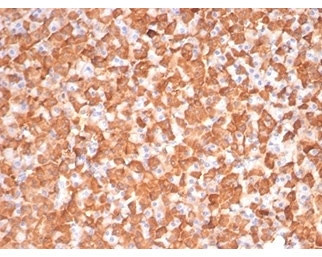
Product information "Anti-PKC iota / PRKCI, clone PRKCI/4911"
1 mg/ml in 1X PBS, BSA free, sodium azide free. Members of the protein kinase C (PKC) family play a key regulatory role in a variety of cellular functions, including cell growth and differentiation, gene expression, hormone secretion and membrane function. PKCs were originally identified as serine/threonine protein kinases whose activity was dependent on calcium and phospholipids. Diacylglycerols (DAG) and tumor promoting phorbol esters bind to and activate PKC. PKCs can be subdivided into at least two major classes, including conventional (c) PKC isoforms (, , , l/i, m and n). Patterns of expression for each PKC isoform differ among tissues and PKC family members exhibit clear differences in their cofactor dependencies. For instance, the kinase activities of PKC ?L and are independent of Ca2+. On the other hand, most of the other PKC members possess phorbol ester-binding activities and kinase activities. Protein function: Calcium- and diacylglycerol-independent serine/ threonine- protein kinase that plays a general protective role against apoptotic stimuli, is involved in NF-kappa-B activation, cell survival, differentiation and polarity, and contributes to the regulation of microtubule dynamics in the early secretory pathway. Is necessary for BCR-ABL oncogene-mediated resistance to apoptotic drug in leukemia cells, protecting leukemia cells against drug-induced apoptosis. In cultured neurons, prevents amyloid beta protein-induced apoptosis by interrupting cell death process at a very early step. In glioblastoma cells, may function downstream of phosphatidylinositol 3-kinase (PI(3)K) and PDPK1 in the promotion of cell survival by phosphorylating and inhibiting the pro-apoptotic factor BAD. Can form a protein complex in non-small cell lung cancer (NSCLC) cells with PARD6A and ECT2 and regulate ECT2 oncogenic activity by phosphorylation, which in turn promotes transformed growth and invasion. In response to nerve growth factor (NGF), acts downstream of SRC to phosphorylate and activate IRAK1, allowing the subsequent activation of NF-kappa-B and neuronal cell survival. Functions in the organization of the apical domain in epithelial cells by phosphorylating EZR. This step is crucial for activation and normal distribution of EZR at the early stages of intestinal epithelial cell differentiation. Forms a protein complex with LLGL1 and PARD6B independently of PARD3 to regulate epithelial cell polarity. Plays a role in microtubule dynamics in the early secretory pathway through interaction with RAB2A and GAPDH and recruitment to vesicular tubular clusters (VTCs). In human coronary artery endothelial cells (HCAEC), is activated by saturated fatty acids and mediates lipid-induced apoptosis. Involved in early synaptic long term potentiation phase in CA1 hippocampal cells and short term memory formation. [The UniProt Consortium]
| Keywords: | Anti-PRKCI, Anti-DXS1179E, Anti-nPKC-iota, EC=2.7.11.13, Anti-PRKC-lambda/iota, Anti-aPKC-lambda/iota, Anti-Protein kinase C iota type, Anti-Atypical protein kinase C-lambda/iota, PKC iota Antibody / PRKCI |
| Supplier: | NSJ Bioreagents |
| Supplier-Nr: | V9171SAF |
Properties
| Application: | IHC (paraffin) |
| Antibody Type: | Monoclonal |
| Clone: | PRKCI/4911 |
| Conjugate: | No |
| Host: | Mouse |
| Species reactivity: | human |
| Immunogen: | A portion of amino acids 100-300 |
| Format: | Ser/Thr Phosphorylation |
Database Information
| KEGG ID : | K06069 | Matching products |
| UniProt ID : | P41743 | Matching products |
| Gene ID : | GeneID 5584 | Matching products |
Handling & Safety
| Storage: | -20°C |
| Shipping: | -20°C (International: -20°C) |
Caution
Our products are for laboratory research use only: Not for administration to humans!
Our products are for laboratory research use only: Not for administration to humans!
Information about the product reference will follow.
more
You will get a certificate here
Viewed