Cookie preferences
This website uses cookies, which are necessary for the technical operation of the website and are always set. Other cookies, which increase the comfort when using this website, are used for direct advertising or to facilitate interaction with other websites and social networks, are only set with your consent.
Configuration
Technically required
These cookies are necessary for the basic functions of the shop.
"Allow all cookies" cookie
"Decline all cookies" cookie
CSRF token
Cookie preferences
Currency change
Customer-specific caching
FACT-Finder tracking
Individual prices
Selected shop
Session
Comfort functions
These cookies are used to make the shopping experience even more appealing, for example for the recognition of the visitor.
Note
Show the facebook fanpage in the right blod sidebar
Statistics & Tracking
Affiliate program
Conversion and usertracking via Google Tag Manager
Track device being used
If you have any questions, please use our Contact Form.
You can also order by e-mail: info@biomol.com
Larger quantity required? Request bulk
You can also order by e-mail: info@biomol.com
Larger quantity required? Request bulk
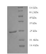
Organism: Homo sapiens (Human). Source: E.coli. Expression Region: 814-1011aa. Protein Length:... more
Product information "Histone deacetylase 9 (HDAC9), partial, human, recombinant"
Organism: Homo sapiens (Human). Source: E.coli. Expression Region: 814-1011aa. Protein Length: Partial. Tag Info: N-terminal 6xHis-tagged. Target Protein Sequence: ILIVDLDVHH GNGTQQAFYA DPSILYISLH RYDEGNFFPG SGAPNEVGTG LGEGYNINIA WTGGLDPPMG DVEYLEAFRT IVKPVAKEFD PDMVLVSAGF DALEGHTPPL GGYKVTAKCF GHLTKQLMTL ADGRVVLALE GGHDLTAICD ASEACVNALL GNELEPLAED ILHQSPNMNA VISLQKIIEI QSMSLKFS. Purity: Greater than 90% as determined by SDS-PAGE. Endotoxin: Not test. Biological Activity: n/a. Form: Liquid or Lyophilized powder. Buffer: If the delivery form is liquid, the default storage buffer is Tris/PBS-based buffer, 5%-50% glycerol. If the delivery form is lyophilized powder, the buffer before lyophilization is Tris/PBS-based buffer, 6% Trehalose, pH 8.0. Reconstitution: We recommend that this vial be briefly centrifuged prior to opening to bring the contents to the bottom. Please reconstitute protein in deionized sterile water to a concentration of 0.1-1.0 mg/mL.We recommend to add 5-50% of glycerol (final concentration) and aliquot for long-term storage at -20 °C/-80 °C. Our default final concentration of glycerol is 50%. Customers could use it as reference. Storage: The shelf life is related to many factors, storage state, buffer ingredients, storage temperature and the stability of the protein itself. Generally, the shelf life of liquid form is 6 months at -20 °C/-80 °C. The shelf life of lyophilized form is 12 months at -20 °C/-80 °C. Notes: Repeated freezing and thawing is not recommended. Store working aliquots at 4 °C for up to one week. Relevance: Binds peptides derived from antigens that access the endocytic route of antigen presenting cells (APC) and presents th on the cell surface for recognition by the CD4 T-cells. The peptide binding cleft accommodates peptides of 10-30 residues. The peptides presented by MHC class II molecules are generated mostly by degradation of proteins that access the endocytic route, where they are processed by lysosomal proteases and other hydrolases. Exogenous antigens that have been endocytosed by the APC are thus readily available for presentation via MHC II molecules, and for this reason this antigen presentation pathway is usually referred to as exogenous. As mbrane proteins on their way to degradation in lysosomes as part of their normal turn-over are also contained in the endosomal/lysosomal compartments, exogenous antigens must compete with those derived from endogenous components. Autophagy is also a source of endogenous peptides, autophagosomes constitutively fuse with MHC class II loading compartments. In addition to APCs, other cells of the gastrointestinal tract, such as epithelial cells, express MHC class II molecules and CD74 and act as APCs, which is an unusual trait of the GI tract. To produce a MHC class II molecule that presents an antigen, three MHC class II molecules (heterodimers of an alpha and a beta chain) associate with a CD74 trimer in the ER to form a heterononamer. Soon after the entry of this complex into the endosomal/lysosomal syst where antigen processing occurs, CD74 undergoes a sequential degradation by various proteases, including CTSS and CTSL, leaving a small fragment termed CLIP (class-II-associated invariant chain peptide). The roval of CLIP is facilitated by HLA-DM via direct binding to the alpha-beta-CLIP complex so that CLIP is released. HLA-DM stabilizes MHC class II molecules until primary high affinity antigenic peptides are bound. The MHC II molecule bound to a peptide is then transported to the cell mbrane surface. In B-cells, the interaction between HLA-DM and MHC class II molecules is regulated by HLA-DO. Primary dendritic cells (DCs) also to express HLA-DO. Lysosomal microenvironment has been implicated in the regulation of antigen loading into MHC II molecules, increased acidification produces increased proteolysis and efficient peptide loading. Reference: Sequence of an HLA-DR alpha-chain cDNA clone and intron-exon organization of the corresponding gene.Lee J.S., Trowsdale J., Travers P.J., Carey J., Grosveld F., Jenkins J., Bodmer W.F.Nature 299:750-752(1982). Function: Responsible for the deacetylation of lysine residues on the N-terminal part of the core histones (H2A, H2B, H3 and H4). Histone deacetylation gives a tag for epigenetic repression and plays an important role in transcriptional regulation, cell cycle progression and developmental events. Represses MEF2-dependent transcription., FUNCTION
| Keywords: | HD9, HD7, HD7b, HDAC7, HDAC9, EC=3.5.1.98, Histone deacetylase 9, Histone deacetylase 7B, Histone deacetylase-related protein, MEF2-interacting transcription repressor MITR, Recombinant Human Histone deacetylase 9 (HDAC9), partial |
| Supplier: | Cusabio |
| Supplier-Nr: | RP179094h |
Properties
| Application: | Activity not tested |
| Conjugate: | No |
| Host: | E.coli |
| Species reactivity: | human |
| MW: | 25.2 kD |
| Purity: | >90% (SDS-PAGE) |
Database Information
| KEGG ID : | K11409 | Matching products |
| UniProt ID : | Q9UKV0 | Matching products |
| Gene ID | GeneID 9734 | Matching products |
Handling & Safety
| Storage: | -20°C |
| Shipping: | +4°C (International: +4°C) |
Caution
Our products are for laboratory research use only: Not for administration to humans!
Our products are for laboratory research use only: Not for administration to humans!
You will get a certificate here
Viewed